A curious guy excited to innovate solutions for a better world.
I recently graduated from my master's at the University of British Columbia (UBC), where I was working on applications of deep learning in medicine. Specifically, I developed novel techniques to make deep learning models more generalizable on pathology images and improve the process of cancer diagnosis. My research interests lie in deep learning, computer vision, and medical images.
Previously, I helped develop a medical device prototype for treating Peyronie's disease (patent application in progress) and worked in pharmaceutical research and production after my bachelor's degree in Chemical and Biological Engineering at UBC.
You can learn more about my career history on my resume.
Select Publications
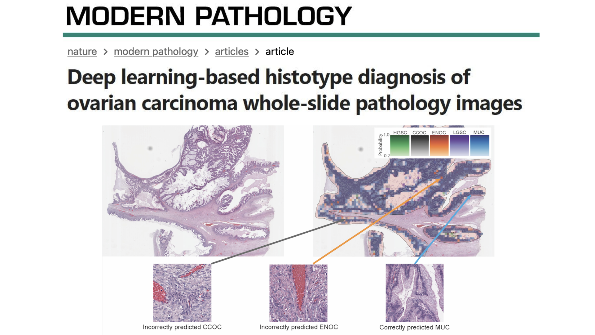
Farahani, H, Boschman, J, et al. “Deep Learning-Based Histotype Diagnosis of Ovarian Carcinoma Whole-Slide Pathology Images.” Modern Pathology, Sept 2022, doi:10.1038/s41379-022-01146-z.
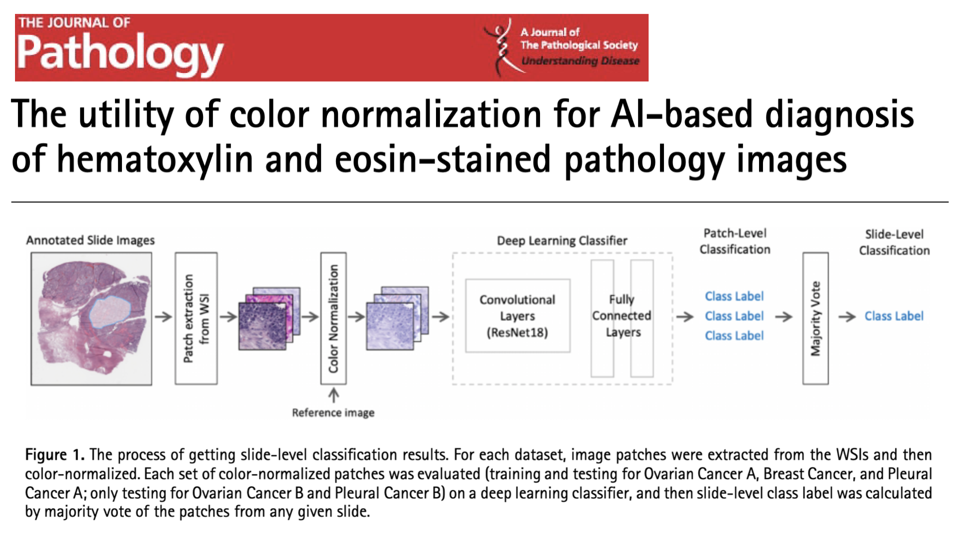
Boschman, J, Farahani, H, et al. “The Utility of Color Normalization for AI-Based Diagnosis of Hematoxylin and Eosin-Stained Pathology Images.” The Journal of Pathology, Sept. 2021, doi:10.1002/PATH.5797.
You can find all my publications through Google Scholar.
Featured Projects

My second, first-author paper is about improving an AI-based ovarian cancer histotype classifier to the performance of expert pathologists and was published in Nature's Modern Pathology. I won multiple awards for this research and it was also featured in this news article.

In my first, first-author paper, we developed a strategy that makes diagnosing digital pathology images with machine learning more generalizable, even when data is sourced from multiple centers.

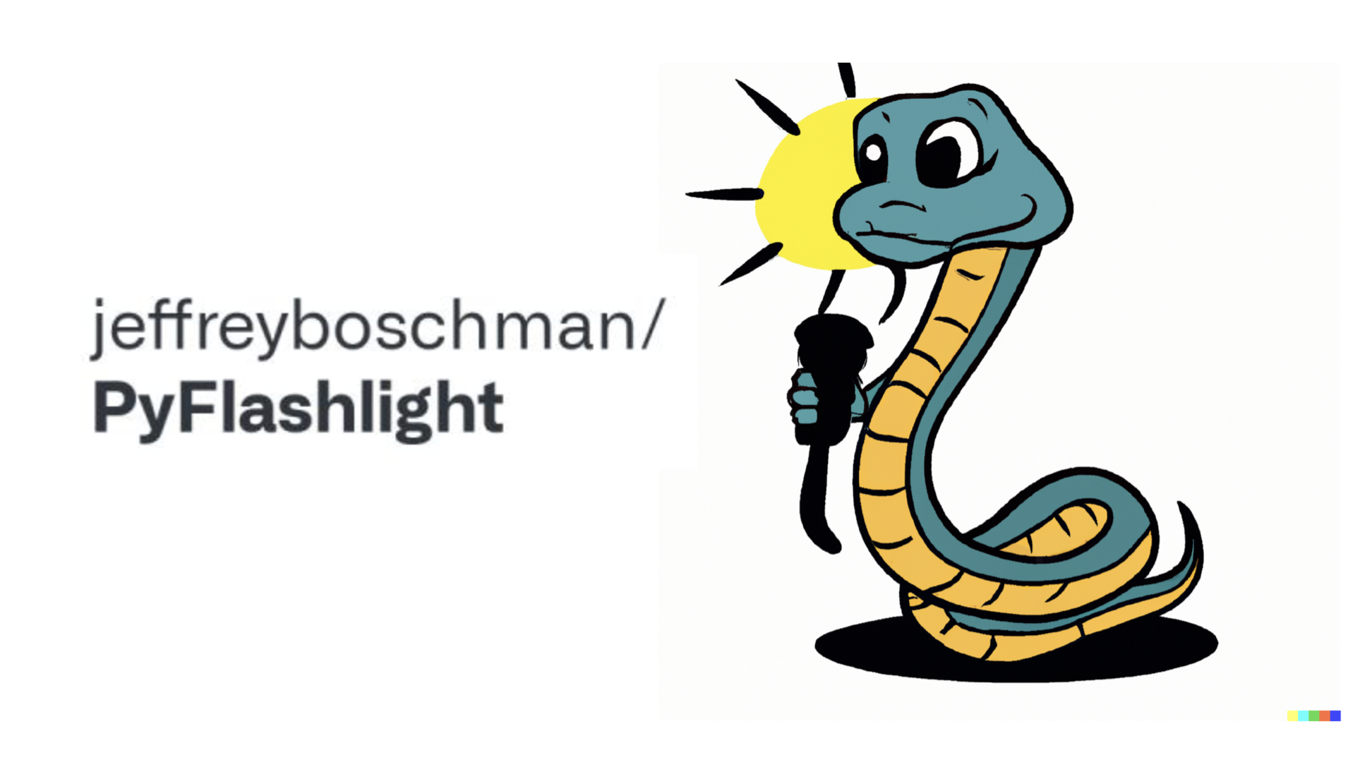
I created PyFlashlight, a Python library to visualize a neural network's computation graph and gain intuition on the backpropagation calculations. It chain together operations, keep track of what operations have been done, and subsequently traverse backwards through them.

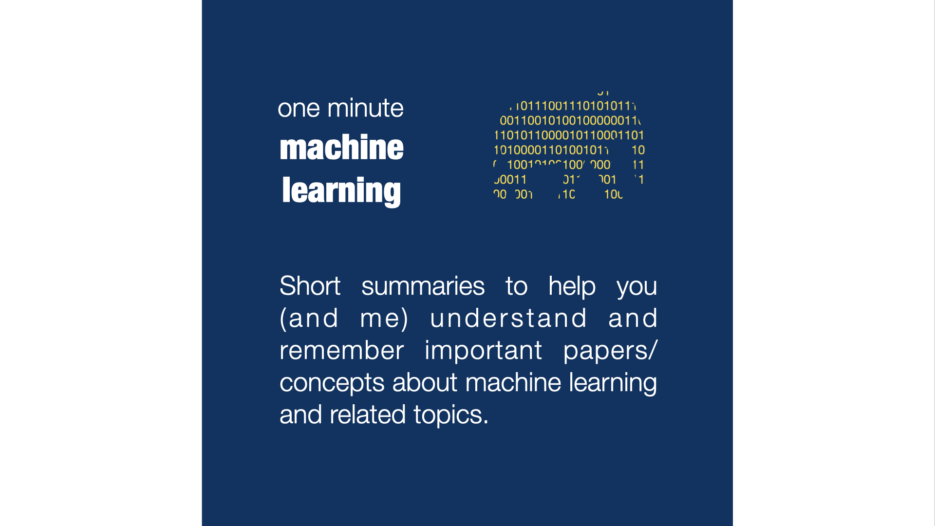
I am the chief author and editor of a publication on Medium dedicated to short articles summarizing important concepts and papers related to machine learning.
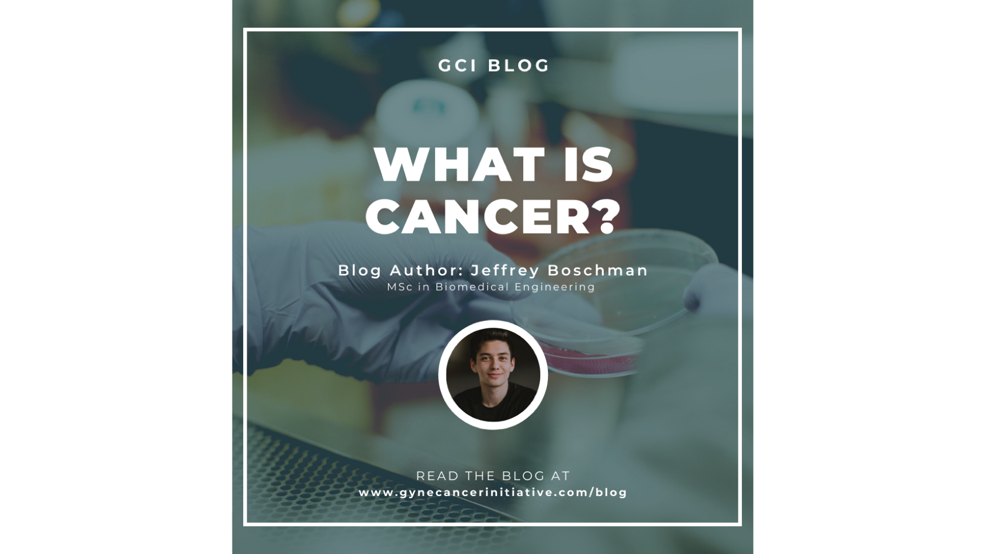
The GCI launched a blog focused on knowledge translation and science communications, and I wrote the first article entitled "What is Cancer?". This platform intends to make information about gynecologic cancers and the gynecologic cancer research in BC more accessible to the general population.
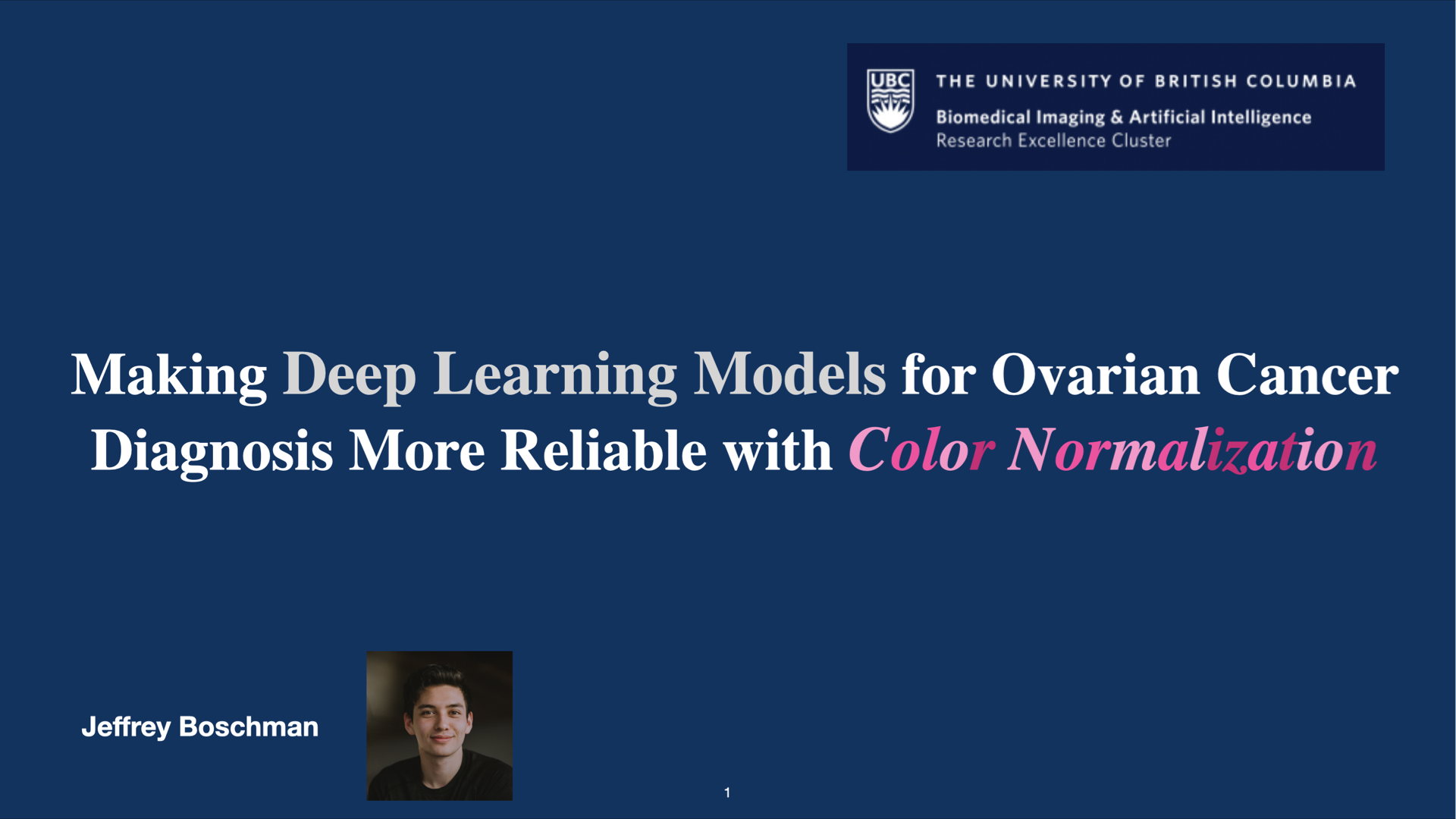
I created a 3-minute video communicating a high-level overview of my research into color normalization for improving deep-learning based classification of histopathology images, which was featured at UBC's Biomedical Imaging and Artificial Intelligence Fall Research Showcase 2021.
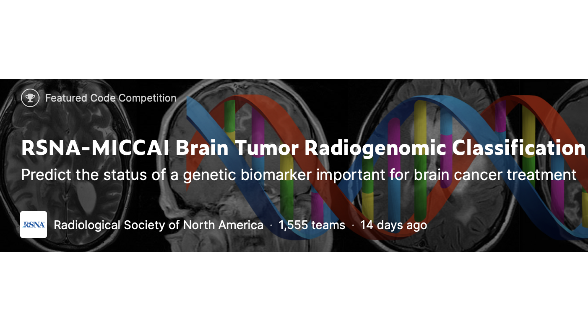
I participated in a Kaggle competition involving predicting whether or not a glioblastoma patient has a certain genetic sequence in their tumor based on 3D MRI DICOM brain scans. This involved working with MRI data (like writing functions to resample data from the sagittal or coronal plane to the axial plane) and creating custom PyTorch models.
Get In Touch
Whether it's an inquiry, constructive feedback, or you just want to chat, I look forward to hearing from you. Please feel free to send an an email or connect with me on LinkedIn.